Release date: 2017-08-30
Recently, the Integrated Cell Materials Research Institute (iCeMS) of Kyoto University in Japan has designed a body-on-a-chip device that can test the side effects of drugs on human cells. The device addresses some of the current problems associated with microfluidic devices and provides new ideas for the next generation of preclinical drug trials. The study was published on the Royal Society of Chemistry Advances on August 24.
Pre-clinical trials of microfluidic devices in place of animals
Nowadays, the development of new pharmaceutical products, including anticancer agents, has many difficulties and requires huge expenses and long-term time. Among these reasons, a particular problem is the pre-clinical trial. In the pre-clinical test before the clinical trial, there are evaluations of the efficacy of the experimental animals, toxicity evaluation tests, and the like. However, it shows that there are many different reactions with humans, so it is difficult to predict the efficacy and toxicity in clinical trials. In addition, the use of experimental animals involves animal protection and is also an ethical issue. Therefore, the development of new test methods has become important to reproduce the reaction of the agent with humans and without animal experiments.
The Kyoto University study used "microfluidic devices" to achieve this goal. Using this device technology, it is possible to mimic the vascular network and tissue in the human body. On this device, cancer cells of human origin and normal cardiomyocytes are carried, and tissues can be connected.
One chip carries two cells that are connected to each other.
This device for detecting heart/cancer on a single chip (iHCC) was used to test the toxicity of the anticancer drug doxorubicin to cardiomyocytes. Researchers led by Ken-ichiro Kamei of iCeMS found that when the drug itself is not heart cytotoxic, the interaction of the drug's metabolites with cancer cells can cause toxicity.
The device is smaller than the microscope slide. It consists of six chambers; each two is connected by a series of inlets and valves, channels. A pneumatic pump controls the flow of fluid in the passage. Each of the two chambers and the respective microchannel system constitutes a test bed. The presence of three test beds in the equipment allows small changes to be introduced in each bed to simultaneously compare the results.
Such a system is more sensitive than cell culture
The team first tested the effects of doxorubicin with separately cultured heart cells and liver cancer cells. The drug has an anticancer effect on cancer cells and does not cause myocardial cell damage.
Then they use the iHCC device to test. Heart cells are placed in one chamber and liver cancer cells are placed in the other. Doxorubicin is introduced into the cell culture medium and circulated through a microchannel closed-loop system that connects the two chambers, mimicking the blood circulatory system. In this way, the drug flows in one direction in a continuous cycle through the two chambers.
The team found signs of toxicity in both cancer cells and heart cells. They speculate that doxorubicinol, a metabolic byproduct of the interaction between doxorubicin and cancer cells, is responsible for the toxic effects.
To test this, they added doxorubicinol to separately cultured heart cells and liver cancer cells and found it to be toxic to heart cells rather than cancer cells.
When doxorubicin is added to liver cancer cells alone, the amount of doxorubicinol produced is too small to be toxic to cardiomyocytes. The team believes this is because the amount of cell culture medium required for the test dilutes the metabolites.
In contrast, when doxorubicin is introduced into iHCC, the flow through the microchannel circulatory system causes the volume of the desired cell culture medium to be small, while the metabolite is not diluted. Therefore, the drug has a toxic effect on cardiomyocytes through its metabolites.
This is a new design concept
The device needs further improvement, but this study suggests that this design concept can be used to study the toxic side effects of anticancer drugs on heart cells before expensive clinical trials. This is the first successful example of connecting multiple organizations on a chip and confirming interactions. This is a model of human in vitro drug mimicry. The function of this new "human body model" device has been confirmed in research and has broad prospects.
Source: Bio-Exploration
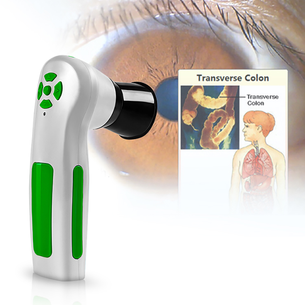
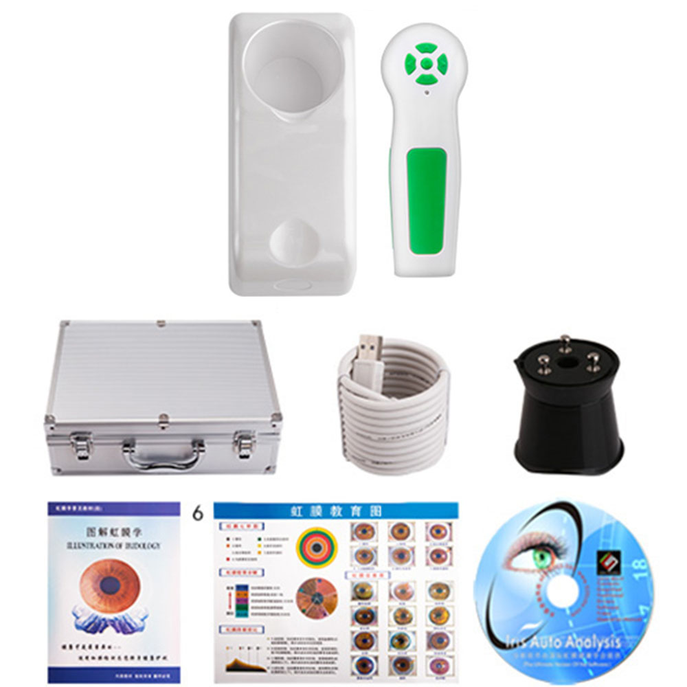
Iridology Camera Health Analyzer
Iris Iriscope Iridology Camera:
Iris holographic diagnostics is a kind of knowledge that applies non-drug and natural therapy to analyze and care for the body by analyzing the iris and pupil to analyze and regulate the health condition of the body. In the field of health medicine, it is called non-invasive health testing and non-drug health intervention. It is a traditional clinical health medicine in Europe.

Iris Iriscope Iridology Camera System Functions:
1. Adjust brightness through either software or switch on handle line, delete the photos, and adjust the focus, quick fixed photos with white balance and high stability of colors. Connects directly with computer without outside power and easy to operate.
2. The software can save the client information and irises also the details of products that you recommend them. After taken the photos, you can analysis the irises and later compare the irises pictures when your client comes back to see their progress. Can print an analysis report. Save the photos according to the date & time you take the photos.3. This machine will help the client know his health condition, including the problem which you have had in the past. The Iridology will be your health counselor, tell you how to keep away from the illness.

Iris Iriscope Iridology Camera Specification:
* Nice appearance and innovative design
* LED illuminator around lens
* Imported lens with plated layer
* 12MP high resolution CCD sensor
* Special DSP image processor, Optical Image Stabilizer
* Single capture button and digital pause capture.
* Adjustable focus to give clear image.
* Auto white balance and contrast adjustment, Color Temperature Filter
* Compatible with iris lens, hair lens.
* Deliver clear and accurate images.
* Easy to operate.
* OS: Windows XP, WIN2000, 2003, Vista, Win7. Win8,win 10
What is the primary benefit of photo iridology?
Not only does it bring clues to your iridology analysis, but it helps to create a better connection with your patient. The patient, who`s naturally curious, is eager to see their own iris. With some simple explanations they can get a basic understanding of your analysis. The connection with your patient strengthens and their understanding of their process improves.
* Iris analysis system: international technology, unique functions.
* Iris analysis system is a medicinal tool that checks the body conditions and prevents diseases from occurring.
* We brought in the advanced iris analysis technology from Germany to lead people to discover sources of illness, and care the body health and spirit in anyways.
* The instrument can show the body conditions of customers and suggest customers the suitable health food, and the plans to care their bodies.
Iridology Camera,Usb Iridology Camera,Iriscope Iridology Camera,Iridology Camera Eye Iriscope
Shenzhen Guangyang Zhongkang Technology Co., Ltd. , https://www.nirlighttherapy.com