Since Roentgen discovered the use of X- rays, live imaging of animals has entered the field of scientists. There are many modes of in vivo imaging, in addition to X- ray ionizing radiation imaging, as well as sound, magnet and even photo-optical imaging.
Each has its own shortcomings and advantages. For example, to determine the location and shape of the anatomy, CT scan, MRI , and ultrasound may be better choices, but they do not provide the injection site and expression level of tumor cells. The necessary information.
Optical imaging: Chemiluminescence and fluorescence technology have been around for decades, and have become an effective method for in vivo imaging due to their relatively low cost, ease of operation, high sensitivity, long-term tracking, and low toxicity.
"The development of fluorescent probes and chemiluminescent reporter technology has made optical imaging widely used in preclinical molecular imaging, " said Simon Cherry of California .
Optical imaging established its potential for drug synthesis in small animal models long before structural imaging was used to detect topological changes. It also intends to establish biomarkers for small animal diseases that will eventually be applied clinically.
Second, the principle of chemiluminescence and fluorescence technology
The principles of chemiluminescence and fluorescence are simple. The former produces light from fireflies and nautical organisms. This is the result of a reaction between luciferase and its substrate, which produces an electronically excited state and then emits light.
Through bioengineering, cells and even animals can produce luciferase, and other like ATP and calcium ions can react similarly, so chemiluminescence becomes an effective tool for many analytical methods.
Fluorescence technology is similar to chemiluminescence except that the fluorescent molecules are excited at different wavelengths and have a shorter lifetime, similar to phosphorescence.
Third, live imaging
Using these principles, promoters, fluorescently labeled antibodies and drugs that drive transgenic luciferase can be imaged in living animals. After injection of intravenous fluorescein substrate or drug, the animal is kept in an opaque chamber, once fluorescent. The enzyme reacts with the substrate and the emitted light is captured by the chemiluminescence detection system. Likewise, fluorescent imaging systems image antibodies or drugs. A sophisticated inspection system and software converts the signals collected from the animal into images for presentation on the screen.
Some imaging systems can simultaneously detect 5 animals and can acquire signals for a long time by continuously inputting anesthetic. The data is dynamic and long-term, for hours, days, or even months.
4. What is the difference between living images from various manufacturers? -- Lips and tongues
However, small animal optical imaging systems are not the same. Some parameters need to be considered. Each system provides its own production, drawing and processing data, but they do very well in some places.
Because there is no external light source and mammals rarely emit chemical light, the background of chemiluminescent imaging systems is very low and their sensitivity is high. But their limitation is that researchers have limited ability to introduce reporters through bioengineering or injection methods, so fluorescence is a better choice.
Fluorescent in vivo imaging requires an external source of light, using a laser or white light source, and the detector placed on the same side of the animal, or opposite.
According to Mark Roskey , vice president of Caliper Life Sciences , the company's IVIS system uses transmitted light illumination, where the detector and light source are opposite the animal and therefore have a significant signal-to-noise ratio.
However, since visible light and a part of near-infrared light are absorbed by the tissue during the detection process, the size of the animal detected by this method is limited. At the same time, more attention should be paid when detecting hemoglobin-intensive organs such as heart, liver and spleen.
The eXplore Optix system developed by Advanced Research Technology (ART) uses one pulse per 100P seconds to excite fluorescence, which not only collects the transmitted wavelength data, but also collects time data. Pierre Couture, vice president of sales and marketing ART Optix think that this life cycle analysis system allows researchers to better understand the process of a particular animal. For example, the decay curve of certain fluorescent molecules changes with changes in the environment, such as pH , or position. This system is several times more expensive than other living images.
 CRI 's systems are renowned in the industry for their combination of electronically tunable filters and multispectral analysis, so they have the advantage of solving autofluorescence or fluorescent crosstalk, enabling simultaneous imaging of multiple fluorescent molecules.
Kodak 's In-Vivo Imaging System FX Pro system provides CT scanning for multimodal imaging , capturing functional (molecular) and structural (anatomical) information on a single machine. " Using our system can do fluorescent, chemiluminescence, and you can see X -ray imaging under the animal with a phosphor screen . "
Zhongke Shengsheng's Molecular Optical Simulation Environment (MOSE) . MOSE is based on the Monte Carlo method to simulate the propagation of photons in biological tissues and free space. The simulation can predict the light intensity signal distribution in the body and surface of small animals.
The advantage of a system, other systems may do the same work in different ways, or do not value the importance of this work.
According to Roskey , IVIS performs multispectral analysis with features such as spectral de- choxing and deconvolution.
Both IVIS and eXplore offer a topological version, and although Couture is somewhat upset, he mentioned, " We have a new version available under Q2 that can do 3D reconstruction. "
Couture also mentioned that eXplore Optix can be used with GE 's eXplore Locus CT scanner to use software to overlay images in three dimensions (however, McLaughlin said Kodak 's software does not require this software).
V. Looking to the future
What are they doing now? What about it later? For example, Kodak and the domestic Zhongke Yusheng are doing research on 3D analysis and multispectral imaging. The latest Micro-CT products introduced by Zhongke Shengsheng have great penetrating power to the tissue, and can be integrated with the fluorescence imaging system to form a multi-modal system, complete animal database, and greatly reduce imaging artifacts.
In general, the industry is addressing the organization of optical imaging that can be used to image dogs, monkeys and even people. Roskey believes that combining brighter, more specific dyes, more advanced bioengineering techniques, and instrument design makes it possible to solve this problem.
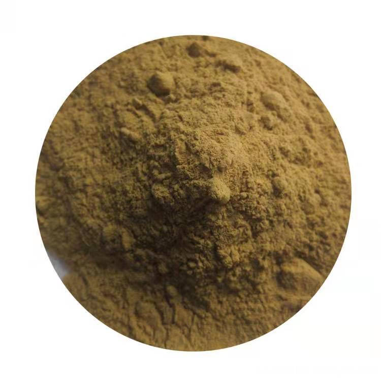
Green Coffee Bean Extract is made from the green beans of the coffea Arabica plant.
There are two types of coffee plants, arabica and robusta. The arabica is higher in quality and higher in chlorogenic and caffeic acids, two primary compounds responsible for anti-oxidant activity. Coffee might have anti-cancer properties, and researchers found that coffee drinkers were 50% less likely to get liver cancer than nondrinkers.
Product features:
1. Special large package for industrial raw material sales(10kg/20kg);
2. 100% pure coffee;
3. Good instant solubility;
4. Stable raw material origin and long-term supply
Functions:
Losing weight.
Anti-virus; Anti-bacteria; Anti-cancer; Anti-aging; Anti-infectious.
Lowering toxicity.
Lowering blood pressure.
Reducing the risk of diabetes.
Help with muscle fatigue for athletes and bodybuilders.
Green coffee bean has strong anti-oxidant properties similar to other natural anti-oxidants like green tea and grape seed extract. Green Coffee Beans have polyphenols which act to help reduce free oxygen radicals in the body. Green Coffee Bean Extract is sometimes standardized to more than 50% Chlorogenic Acid. Chlorogenic Acid is the compound present in coffee which has been long known as for its beneficial properties. This active ingredient makes green coffee bean an excellent agent to absorb free oxygen radicals; As well as helping to avert hydroxyl radicals, both which contribute to degradation of cells in the body.

Coffee Green Hydrochloric Acid
Coffee Bean Extract,Robusta Coffee Extract,Pure Green Coffee Bean Extract,Coffee Green Hydrochloric Acid
Yunnan New Biology Culture Co,.Ltd , https://www.lvsancoffee.com